Tea vs Alzheimer’s Disease

A life of hardship has left only the slightest marks on her hands, which now clinch to one another. Maybe that is the only thing that she can physically hold tightly on to. Everything else is slipping away.
 I have a confession to make: I have not been able to make my mother drink tea regularly. In fact, she is slight even for water. My mother has an Alzheimer’s condition that is worsening. Sometimes I blame myself for failing to make tea her habit years earlier. Seeing her memory fading away had been mixed feelings of sadness, regrets and guilt. It is only very recently that I have learned to accept it as part of her passage of life and embrace her gradual mental backward evolvement into a small child.
For the readers, however, I am presenting some recent findings and how I read these as the relationship between tea and neurological health. Hopefully this writing can help some lucky ones to prevent Alzheimer’s from happening, or at least to a lesser extent or until much later in life.
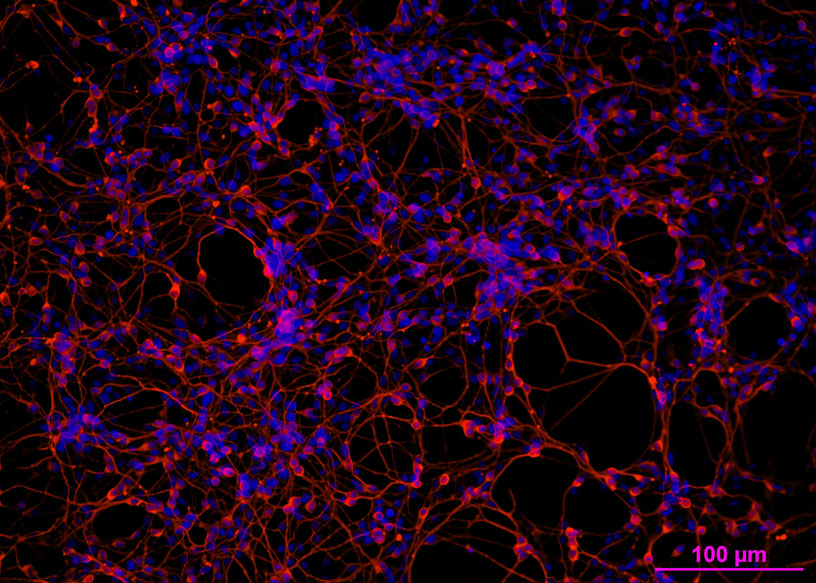
Closeup of a part of a network of neuron cells. The nervous system, including the whole brain, is composed of complex multi-networks made up of neurons reaching out to one another with long filament structures, just like wires and plugs.
What is Alzheimer’s Disease?
Forgetfulness, the symptom that we all know Alzheimer’s Disease (AD) of, is only but one of the sufferings of the disease.
The disease is basically a degeneration of the brain. W.H.O. has another name for it: the Brain Killer. The neuron cells in the brain get deformed and die. The connections between them, which are the basis to our brain functions, are gradually cut off. Slowly as the brain shrinks, the patient loses her cognitive ability, becomes agitated easily, has depression, delusions, and/or hallucinations. She is more and more withdrawn from her surrounding, including her immediate family. Increasingly she has no control over her bladder and bowels, and also her motor and verbal skills. This can get to a point when she cannot even control her muscles to swallow properly. As the disease progresses, it becomes lethal.
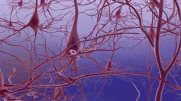
Destruction of the brain, one neuron at a time

A structural diagram of a neuron cell
The big fat red structure is the main cell body. It reaches out with long branches that are called axons, the axons further branch out in dendrites with endings that form synapses — the plugs and connections of the nervous system. For a comprehensive caption, see below »
The surface of our brain is made up with neuron cells. Their normal functionalities, including their interconnectivity, allow a person to think, perceive, remember, stay conscious and to control speech and movement.
The neuron cell reaches out to connect with one another by branching out with tentacle-like endings. Signals are passed through these endings across gaps called synapses. Our daily activities are made possible by the trillions of sending and receiving signals across these synapses.
A particular kind of protein molecule, “tau” protein, that normally plays a contributive part in the function of these synapses deforms and disintegrates in much larger quantity than normal in patients with Alzheimer’s. These filament-like micro-tube molecules disintegrate and tangle with one another within the neuron cell — what the science community call neurofibrillary tangles — and become toxic. This toxicity gradually makes the cell kill itself.
Protein is messed up not only within the cell, but also outside of it. Clusters of protein structure — beta amyloid (Aß) — aggregate in between the neurons, forming plaques that interfere with signal transmission, causing inflammation, cutting off the synapses, triggering a series of bad chemical actions and ultimately deaths of neurons. That is why W.H.O. has another name for the disease: Brain Killer. Aß is now seen as a major target in combating Alzheimer’s Disease.
Neuronal death and the formation of neurofibrillary tangles and beta-amyloid plaques. Animation credit: National Institute on Aging
Alzheimer’s is lethal
While some patients may live on 20 years more, most die between 4 to 8 years after diagnosis. There is no established medical prevention or cure for the disease. As the medical and scientific communities work hard collectively for the improvement of our survival against most other fatal ailments, Alzheimer’s Disease is the only major cause of deaths with increasing fatal rate, compared with HIV, circulatory diseases or even cancers.
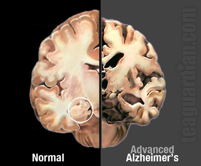
Comparing the autopsy dissections of the brain of an Alzheimer’s disease patient and that of a normal person reveals how much the brain of an AD patient has shrunk. Notice in particular the Hippocampus — the part of the brain that is critical in consolidation of information, including memory and spatial perception, and that is circled in white in the normal brain — has virtually disappeared in the patient’s.
W.H.O. has another name for it: the Brain Killer
Another threat of the disease is the cost of care-taking the patient. A person who has lost her memory and ability to understand her environment is in danger even in her own home.
My mother used to live with one of my sisters. Before we confirmed that she had Alzheimer’s, she herself had taken on hiding in the apartment and afraid to go out. I went there once or twice a week during the day when my sister was at work so I could take her out for a walk in the park and some simple exercises. As conditions worsened my sister hired a maid to look after her. It was a total nightmare and ended almost in tragedy. I think both my mother and the maid were not prepared for the rapid development of the disease and what it really means in behavioural patterns.
caption to “A structural diagram of a neuron cell”:
- Rough ER ( Nissi body )
- Polyribosomes
- Ribosomes
- Golgi aparatus
- Nucleus
- Nucleolus
- Membrane
- Microtubes
- Mitochondrion
- Smooth ER
- Axon hillock
- Nucleus ( Schwann cell )
- –
- Synapse
- Dendrites
- Axon
- Neurontransmitter
- Receptor
- Synapse closeup
- Microfilaments
- Myeline Sheath ( Schwann cell )
- Node of Ranvier
- Axonal terminal
- Synaptic vesicles
- Synapse ( Axoaxonic )
- Synaptic cleft